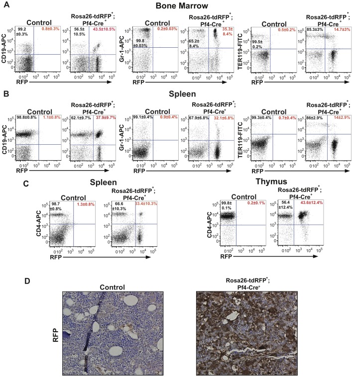
Figure 2. Pf4-Cre activates RFP expression in all hematopoietic lineages.
(A) Frequencies of RFP-expressing B-lymphoid (CD19+), myeloid (Gr-1+), and erythroid (Ter119+) cells in the bone marrow obtained from adult Rosa26-tdRFP+;Pf4-Cre+ mice and Rosa26-tdRFP+;Pf4-Cre− litter matched controls. (B) Frequencies of RFP-expressing B-lymphoid (CD19+), myeloid (Gr-1+), and erythroid (Ter119+) cells in the spleen from adult Rosa26-tdRFP+;Pf4-Cre+ mice and Rosa26-tdRFP+;Pf4-Cre− litter matched controls. (C) Frequencies of RFP-expressing CD4+ T cells in the spleen and thymus of adult Rosa26-tdRFP+;Pf4-Cre+ mice and Rosa26-tdRFP+;Pf4-Cre− litter matched controls. Dot plots shown in Figure 1A–C are representative figures of 3–5 independent experiments with values shown as an average±SEM from 3–5 independent experiments. % of RFP+ cells is indicated in red and % of RFP− in black. (D) Bone marrow from Rosa26-tdRFP+;Pf4-Cre+ and litter matched controls was fixed, and histologically stained for RFP. Images are representative of 2 independent experiments.

