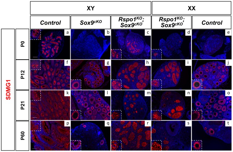
Figure 3. Post-natal development of sex cords in XY and XX Rspo1KOSox9cKO mice.
Immunofluorescence of SDMG1 (in red). Counterstain is DAPI (in blue). SDMG1 is expressed in Sertoli cells (XY controls a, f, k, p) and in follicular cells of growing ovaries as evidenced at P12 onwards (j, o, t). Sertoli cells are present and formed sex cords in both XY and XX Rspo1KOSox9cKO gonads, with more developing sex cords in XY Rspo1KOSox9cKO testis (c, h, m, r) in comparison to XX Rspo1KOSox9cKO ovotestis (d, i, n, s). At P12, the sex cords are fully developed in both XY (h) and XX (i) Rspo1KOSox9cKO mice. In XY Sox9cKO (b, g, l, q) and XX control (e, j, o, t) gonads, ovarian follicles express SDMG1 at P12, P21 and P60. At these stages, SDMG1 is also expressed in the follicles of the XX double mutant ovotestes (see n) and in XY double mutant follicles when they develop (scale bars: 100 µm). XY (a, f, k, p) and XX (e, j, o, t) Rspo1+/−; Sox9flox/flox controls, XY Sox9cKO gonads (b, g, l, q), XY (c, h, m, r) and XX (d, i, n, s) Rspo1KO Sox9cKO gonads respectively.

