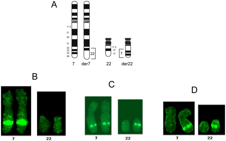
Figure 3. Representative fluorescent in situ hybridization (FISH) patterns of DNA probes onto chromosomes 7, 22 der(7) and der(22) from this patient’s chromosome spreads.
(A) Schematic drawing of normal chromosomes 7 and 22 and derivative chromosomes der(7) and der(22), showing the translocated chromosomal regions. The three panels below depict each the typical hybridization of a probe located (B) on chromosomes 7, der(7), (C) on chromosomes 7, der(7), der(22) and (D) chromosomes 7 and der(22).

