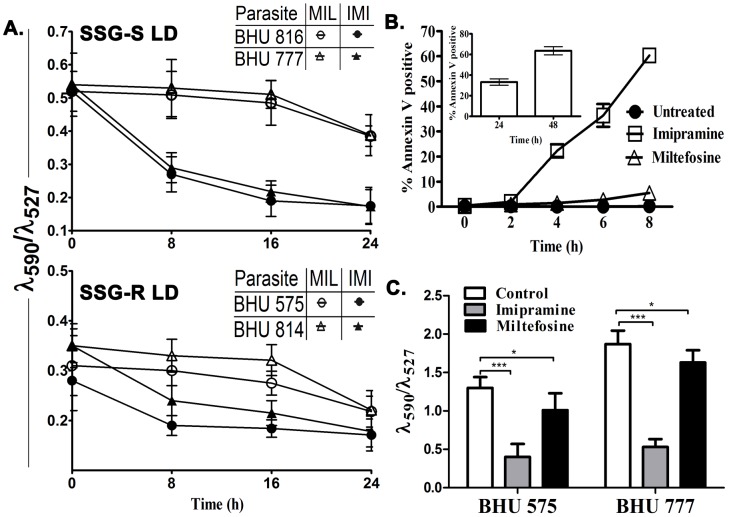
Figure 1. Imipramine and miltefosine induced alteration of mitochondrial transmembrane potential and apoptosis induction in LD promastigotes.
A. BHU 816(S), BHU 777(S), BHU 575(R) and BHU 814(R) promastigotes were exposed to 75 µM imipramine (•,▴) or 40 µM miltefosine (○,Δ) for 8, 16 and 24 h and the mitochondrial transmembrane potential was determined using JC-1 fluorescent probe. The fluorescence was measured at 527 and 590 nm and the ratio (λ590/λ527) was plotted. Three independent experiments were performed and mean±SE was presented. B. Apoptosis of LD parasites as a measure of % Annexin V positive cells in BHU 575(R) in the presence of 75 µM imipramine (□) or 40 µM miltefosine (Δ) and absence of drug (•) was measured as a function of time. Phosphatidylserine exposure was analyzed by flow cytometry. Inset 1B showing the percentage of apoptotic cells in BHU 575(R) upon miltefosine (40 µM) treatment at 24 and 48 hour. The data are the mean of three independent experiments, and standard deviations are represented by error bars. C. BHU 777(S) and BHU 575(R) amastigotes were subjected to either 75 µM imipramine or 40 µM miltefosine treatments for 8 h and the resulting mitochondrial transmembrane potential was measured using JC-1 as above.

