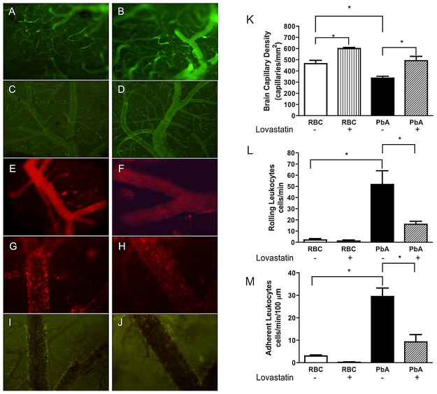
Figure 4. Lovastatin improves microvascular function and decreases leukocyte rolling and adhesion during PbA infection.
A–J: representative images of fluorescence intravital microscopy of pial vessels of uninfected mice (A and B; Magnification 100×) or infected mice (C and D) treated with vehicle or lovastatin, respectively (Magnification 40×). Venules with adherent and rolling rhodamine-labeled leukocytes in uninfected mice (E and F) or infected mice (G and H) treated with vehicle or lovastatin, respectively (Magnification 200×). Rhodamine-labeled leukocytes were associated with the venular endothelium in animals treated with vehicle (G) whereas they were largely free in the blood stream in lovastatin-treated mice (H). Fluorescent Plasmodium berghei (GFP)-infected RBC in venules of mice treated with vehicle (I) or lovastatin (J) (Magnification 200×). Mean ± SEM of functional capillary density (K), of rolling-rhodamine labeled leukocytes (L), and of number of adherent leukocytes (M) in pial venules (n = 6/group); *p<0.05 in relation to control (RBC) and vehicle-treated groups (Bonferroni's Multiple Comparison Test) and between PbA and PbA-lovastatin group (Bonferroni's Multiple Comparison Test; Scale bar, 100 µm).

