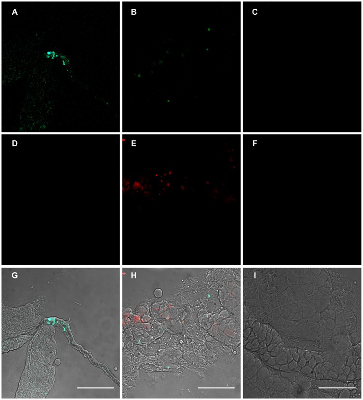
Figure 5. Localization of Wolbachia and DENV-2 in Ae. albopictus.
At 14 days post-DENV-2 infection, salivary glands were dissected, fixed, and then incubated simultaneously with two Wolbachia probes and two DENV specific probes. In panels A, B, and C, DENV-2 (green) is labeled with FITC. In panels D, E, and F, Wolbachia (red) is stained with Rhodamine. In panels G, H, and I, the red and green channels are merged. A co-localization of Wolbachia and DENV-2 was detected in some cells (panel H). DENV-uninfected and Wolbachia-uninfected controls are presented in panel C and F, respectively. Scale bars: 50 µm.

