
Figure 7.
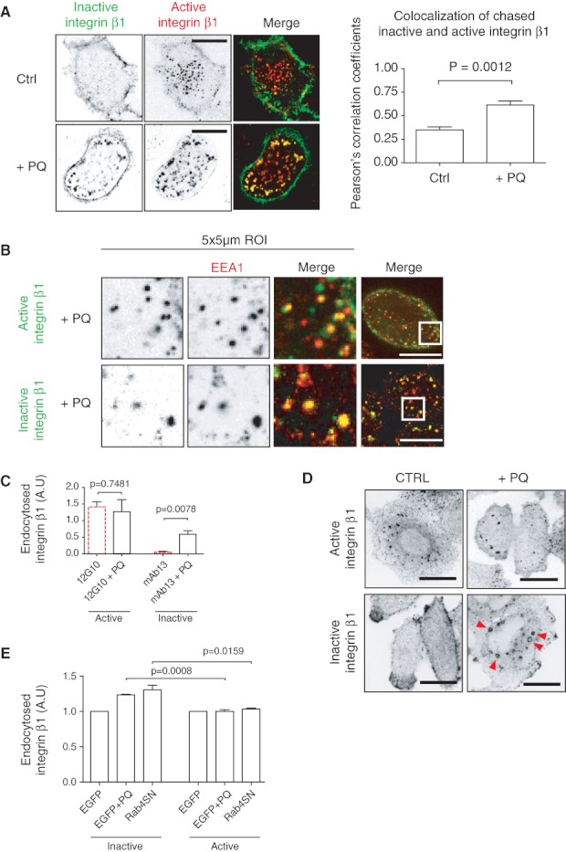
Inhibition of recycling increases the amount of endocytosed inactive β1 integrins. A) NCI-H460 cells were double labelled for 1 h on ice with β1 integrin antibodies against active (12G10) and inactive (mAb13) conformations. To block the recycling, 0.5 m m PQ was added during the 30-min endocytosis at +37°C. Cells were fixed and imaged with confocal microscope. Scale bar 10 µm. Columns show mean + standard error of the mean (SEM) of PC coefficients of the colocalization over the whole image between active and inactive β1 integrins (n = 10). p Values are calculated using Mann–Whitney test. B) NCI-H460 cells treated with 0.5 m m PQ were allowed to endocytose antibodies against active (12G10) and inactive (mAb13) β1 integrins. Cells were fixed, permeabilized and stained against early endosome marker EEA1. Scale bar 10 µm. C) Results of antibody-based endocytosis assay using fluorescent plate-reader. NCI-H460 cells were allowed to endocytose active (12G10) and inactive (mAb13) β1 integrin with and without 0.5 m m PQ for 30 min at 37°C. The graphs show mean + SEM of three independent experiments. p Values are calculated using Mann–Whitney test. D) NCI-H460 cells were treated with 0.5 m m PQ for 30 min at 37°C, fixed and stained against active (12G10) and inactive (mAb13) integrin β1. Arrowheads point to endosomes containing inactive β1 integrin. E) MDA-MB-231 cells were transfected with EGFP-alone or EGFP-Rab4a-S22N. Cells were harvested with HyQtase and surface stained against inactive (mAb13) or active (12G10) β1 integrins. Integrins were allowed to endocytose for 60 min, and the level of cell surface β1 integrin was analysed from non-treated, PQ-treated and EGFP-Rab4a-S22N positive cells before and after endocytosis. Columns show level of endocytosed β1 integrin normalized to EGFP-control, mean + SEM of three independent experiments. p Values are calculated using unpaired t-test.