
Figure 8.
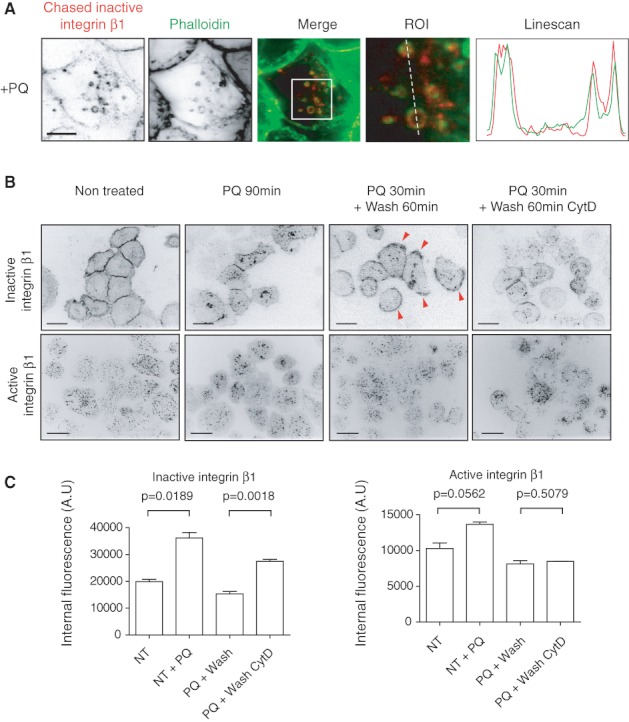
Inactive β1 integrin recycling is dependent on actin. A) NCI-H 460 cells were labelled for 1 h on ice and allowed to endocytose inactive β1 integrin ( mAb 13, A lexa F luor 647-labelled) for 30 min at 37°C with 0.5 m m PQ. Cells were fixed and stained with A lexa F luor 488 P halloidin to visualize F -actin. A line scan and ROI are shown on the right. Scale bar 10 µm. B) NCI-H 460 cells were surface labelled against inactive ( mAb 13) or active (12 G1 0) β1 integrin for 1 h on ice and incubated at 37°C with growth medium (non-treated) for 90 min, with 0.5 m m PQ for 90 min, with 0.5 m m PQ for 30 min followed by a 60-min medium wash, with 0.5 m m PQ for 30 min followed by a 60-min wash with medium containing 20 m m CytD. Mid-slice confocal images are shown. Arrowheads point to membrane-relocalized inactive β1 integrin. Scale bar 10 µm. C) NCI-H 460 cells were surface labelled against inactive ( mAb 13) or active (12 G1 0) β1 integrin 60 min on ice and incubated at 37°C with growth medium (non-treated) for 60 min, with 0.5 m m PQ for 60 min, with 0.5 m m PQ for 30 min followed by a 30-min medium wash, with 0.5 m m PQ for 30 min followed by a 30 min wash with medium containing 20 m m CytD. Cells were lifted on ice and cell surface fluorescence was quenched. The level of internal β1 integrin was measured using automated fluorescent microscope ScanR. Columns show mean + standard error of the mean of 6000–8000 cells. P V alues are calculated using unpaired t-test.