
Figure 9.
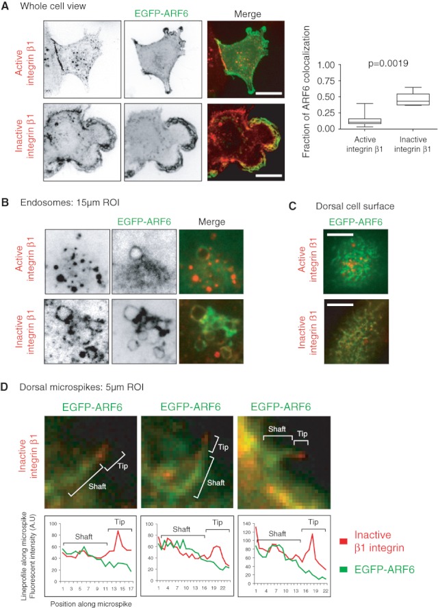
Inactive β1 integrin localizes to ARF6-positive endosomes and protrusions. MDA-MB-231 cells were transfected with EGFP-ARF6. Cells were surface labelled against inactive (mAb13) or active (12G10) β1 integrin. Integrins were allowed to endocytose 120 min, cells were fixed, counterstained and analysed under confocal microscope. A) Mid-sections are shown. Scale bar 10 µm. The fraction of ARF6 colocalization with active and inactive β1 integrin was quantified. Graph shows mean ± standard error of the mean. p Value was calculated using Mann–Whitney test. B) Fifteen micrometre ROIs of ARF6-positive endosomes are shown. C) Dorsal cell surfaces are shown. Scale bar 10 µm. D) Five micrometre ROIs of dorsal cell surface are shown with linescan along the ARF6-positive microspike. Shaft and tip regions are illustrated over the ROI and the corresponding region marked over the linescan.