Figure 7.
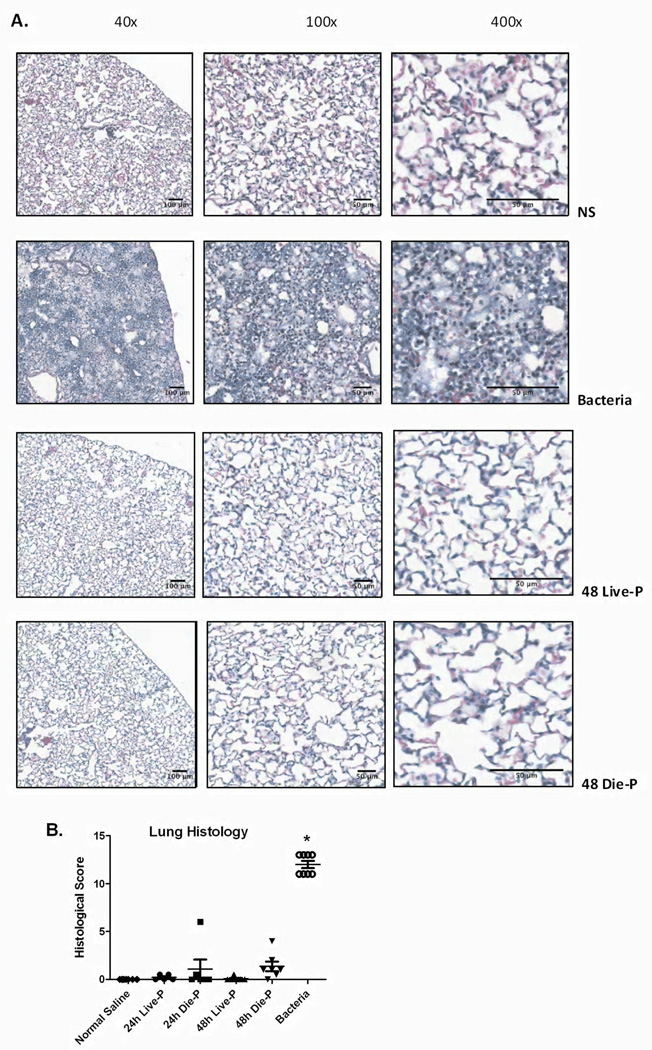
Histopathological evaluation of the lung 24 and 48 hours after cecal ligation and puncture. In Figure 7 A, images of hematoxylin and eosin stained slides at increasing magnifications are shown for 48 hour Die-P and Live-P mice, as well as a mouse given normal saline or bacteria. No appreciable signs of edema, protein leakage or alveolar neutrophil infiltration could be detected at any magnification in either Live-P or Die-P mice. Histological scoring is shown for each group in Figure 7 B (24h Live-P (n=5), 24h Die-P (n=5); 48h Live-P (n=6); 48h Die-P (n=7), normal saline (n=8), bacteria (n=8). The sections were evaluated in a blinded manner by a board certified pathologist. Values are mean ± SEM. There were significant differences between all the groups, p<0.001, but there was no difference between Live-P and Die-P mice at either 24 or 48 hours. * = p<.05 for bacteria versus normal saline.

