
Figure 1.
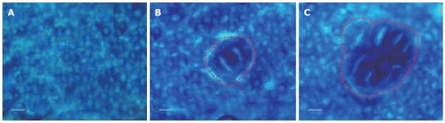
Topographical view of colon tissue. A: Normal colon tissue; B: Aberrant crypt foci (ACF) with 2 crypts; C: Large ACF (crypt multiplicity containing more than 3 crypts per ACF). Scale bars = 25 μm.
Official websites use .gov
A
.gov website belongs to an official
government organization in the United States.
Secure .gov websites use HTTPS
A lock (
) or https:// means you've safely
connected to the .gov website. Share sensitive
information only on official, secure websites.
Topographical view of colon tissue. A: Normal colon tissue; B: Aberrant crypt foci (ACF) with 2 crypts; C: Large ACF (crypt multiplicity containing more than 3 crypts per ACF). Scale bars = 25 μm.