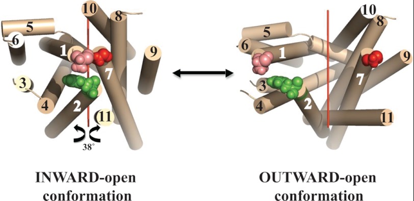
FIGURE 1.
Ab initio computational model of LdNT1.1. Left, indicated are residues that were mutated to cysteines. Tan cylinders represent predicted TM helices and are numbered 1–11. Residues at the extracellular termini of helices 1, 2, and 7 that were mutated to cysteines are indicated by space filling models: pink is Ala-61TM1, green is Phe-74TM2, and red is Gly-350TM7. The view is from the extracellular surface toward the interior, indicating that the ab initio model predicted an inward-open conformation. The figure was generated using PyMol. A suggestive model for the outward-open conformation (right) is given by rotating the N-terminal domain (helices 1–6) and the C-terminal domain (helices 7–11) 38° around an axis (red line) parallel to the lipid bilayer, as explained in “Results.”

