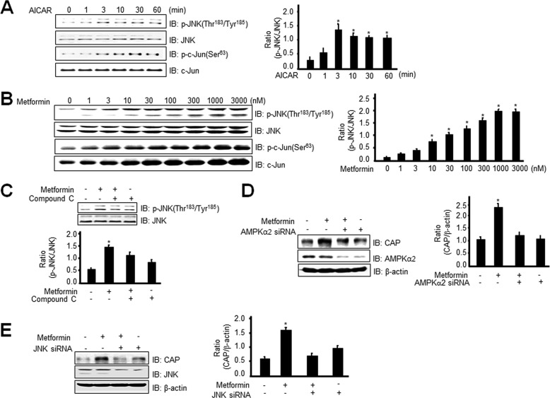
FIGURE 4.
Metformin stimulates CAP expression through phosphorylation of JNK in an AMPK-dependent manner. A, 3T3-L1 preadipocytes were stimulated for the indicated times with 1 mm AICAR. Cell lysates (25 μg) were analyzed via Western blotting (IB) with an anti-phospho-JNK (Thr183/Tyr185) or anti-phospho c-Jun (Ser63) antibody. Blotting with the anti-JNK or c-Jun antibody was used as a protein-loading control. Data in the bar graphs represent the mean ± S.E. (error bars) values of the ratios of densities (p-JNK/JNK) for at least three individual Western blotting experiments. *, p < 0.05 versus basal values. B, 3T3-L1 preadipocytes were stimulated with the indicated doses of metformin for 6 h. Cell lysates (25 μg) were analyzed via Western blotting with an anti-phospho-JNK (Thr183/Tyr185) antibody. Blotting with the anti-JNK antibody was used as a protein-loading control. Data in the bar graphs represent the mean ± S.E. values of the ratios of densities (p-JNK/JNK) for at least three individual Western blotting experiments. *, p < 0.05 versus basal values. C, 3T3-Ll preadipocytes were stimulated for 30 min with 1 mm metformin in the presence of compound C. Cell lysates (25 μg) were analyzed via Western blotting with an anti-phospho-JNK (Thr183/Tyr185) antibody. Blotting with the anti-JNK antibody was used as a protein-loading control. Data in the bar graphs represent the mean ± S.E. values of the ratios of densities (p-JNK/JNK) for at least three individual Western blotting experiments. *, p < 0.05 versus basal values. D, 3T3-L1 preadipocytes were transiently transfected with AMPKα2 siRNA for 48 h, prior to 1 mm metformin treatment for 6 h. Cell lysates (25 μg) were analyzed via Western blotting using the anti-CAP or AMPKα2 antibody. Blotting with the anti-β-actin antibody was used as a protein-loading control. Data in the bar graphs represent the mean ± S.E. values of the ratios of densities (CAP/β-actin) for at least three individual Western blotting experiments. *, p < 0.05 versus basal values. E, 3T3-L1 preadipocytes were transiently transfected with JNK siRNA for 48 h, prior to 1 mm metformin treatment for 6 h. Cell lysates (25 μg) were analyzed via Western blotting with the anti-CAP or JNK antibody. Blotting with anti-β-actin antibody was used as a protein-loading control. Data in the bar graphs represent the mean ± S.E. values of the ratios of densities (CAP/β-actin) for at least three individual Western blot experiments. *, p < 0.05 versus basal values.

