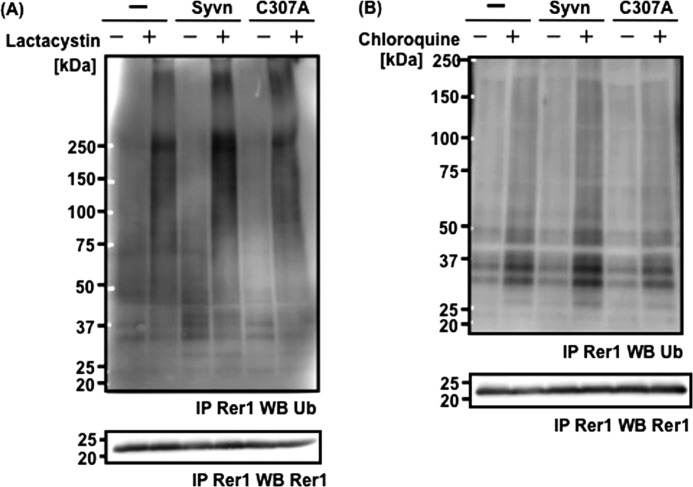
FIGURE 7.
Rer1 is ubiquitinated by Syvn. A, Rer1 immunoprecipitation from the following cell lines treated with 10 μm lactacystin for 10 h (lower panel): Syvn−/− fibroblasts infected with pMX (-), pMX-Syvn-FLAG (Syvn), or pMX-C307A-FLAG (C307A) together with pMX-hRer1. Ubiquitinated Rer1 precipitated by the anti-Rer1 antibody was detected by Western blotting with anti-mono- and polyubiquitin antibody (upper panel). B, Rer1 was immunoprecipitated from Syvn−/− fibroblasts treated with 50 μm chloroquine for 24 h (lower panel) and infected as shown in Fig. 7A. Ubiquitinated Rer1 precipitated with the anti-Rer1 antibody was detected by Western blotting with anti- mono- and polyubiquitin antibody (upper panel).

