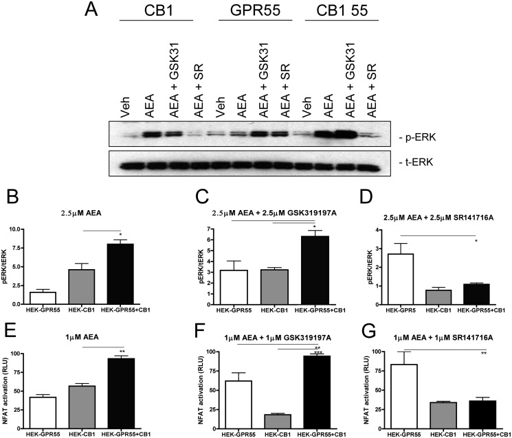
FIGURE 6.
Combined administration of the endogenous CB1 agonist anandamide and GPR55 ligands restore GPR55-mediated ERK1/2 phosphorylation and NFAT activation in HEK-GPR55+CB1 cells. Representative immunoblot showing ERK1/2 phosphorylation in response to vehicle, 2.5 μm AEA, 2.5 μm AEA + 2.5 μm GSK319197A and 2.5 μm AEA + 2.5 μm SR141716A in HEK-GPR55, HEK-CB1, and HEK-GPR55+CB1 cells (A). Activation of ERK1/2 (A-D) and NFAT (E–G) were altered in HEK-GPR55, HEK-CB1, and HEK-GPR55+CB1 cells after individual stimulation with 2.5 μm AEA (A, B, and E) or co-stimulation with 2.5 μm AEA + 2.5 μm GSK319197A (A, C, and F) or 2.5 μm AEA + 2.5 μm SR141716A (A, D, and G). Stimulation with 2.5 μm AEA leads to a significantly higher level of ERK1/2 (A and B) and NFAT (E) activation in HEK-GPR55+CB1 compared with HEK-CB1 cells. No activation over baseline was observed in HEK-GPR55 cells. Increased ERK1/2 phosphorylation (A and C) and NFAT (F) activation occurred in all three cell lines after co-stimulation with 2.5 μm AEA and 2.5 μm GSK319197A. HEK-GPR55+CB1 cells show significantly higher activation levels compared with single cell lines. pERK1/2 (A and D) and NFAT (G) activation is inhibited by co-stimulation of HEK-CB1 and HEK-GPR55+CB1 cells with 2.5 μm AEA and 2.5 μm SR141716A, but induced in HEK-GPR55 cells. pERK1/2 was normalized to total ERK1/2 and data are means of three independent experiments ± S.E. Reporter gene assay data are mean ± S.E. from one of four independent experiments performed in triplicates. Data were normalized and expressed as percent of maximum activation, which was set as 100%, *, p < 0.05; **, p < 0.01; ***, p < 0.001.

