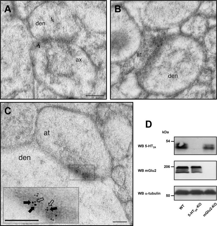
FIGURE 1.
Subcellular co-localization of 5-HT2A and mGlu2 receptors in mouse cortical neurons. A, immunogold labeling for the 5-HT2A receptor. B, immunogold labeling for the mGlu2 receptor. C, immunogold labeling for 5-HT2A and mGlu2 receptors. Different sized black dots show 5-HT2A receptor immunolabeling (larger 10-nm gold particles) and mGlu2 receptor immunolabeling (smaller 6-nm gold particles). Inset, high magnification view of region delineated by boxed area. Note that the 10-nm gold particles (filled arrows) and the 6-nm gold particles (open arrows) are located in very close proximity at synaptic junctions in mouse frontal cortex (den, dendrite; ax, axon; at, axon terminal). Scale bars, 100 nm. D, Western blots (WB) in frontal cortex of wild type (WT), 5-HT2A-KO, and mGlu2-KO mice. Representative immunoblots in plasma membrane preparations of mouse frontal cortex are shown.

