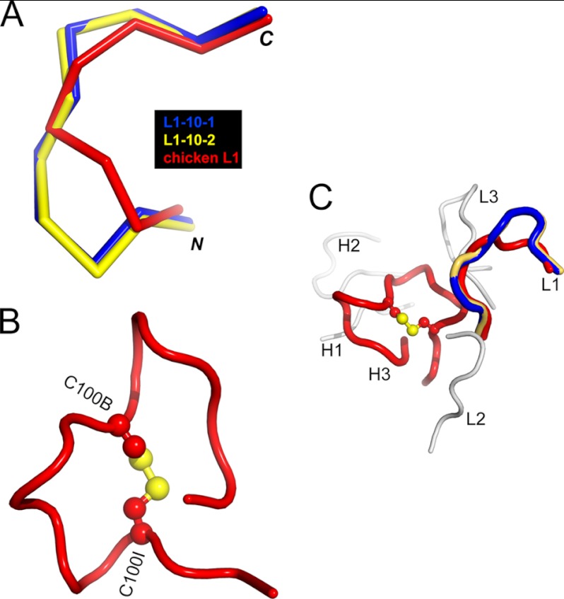
FIGURE 5.
Chicken-specific conformations of CDR-L1 and CDR-H3 from anti-phospho-tau Fab (pT231/pS235_1). A, ribbon view showing the superposition of chicken CDR-L1 (red) with canonical mammalian CDR-L1–10-1 (blue; Protein Data Bank code 1YQV) and CDR-L1–10-2 (yellow; code 1AY1), respectively. CDR-L1–10-1 and CDR-L1–10-2 represent two clusters of mammalian CDR-L1 conformations and show the best structural similarity to chicken CDR-L1. B, schematic view showing the conformation of chicken CDR-H3 stabilized by the intramolecular disulfide bond shown as a ball and stick model in atomic colors (carbon, red; and sulfur, yellow). Two cysteines are labeled C100B and C100I, respectively. C, schematic view showing chicken CDRs with the same coloring scheme as in A and B. CDR-L2, CDR-L3, CDR-H1, and CDR-H2 are shown in gray.

