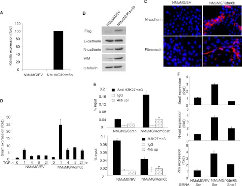
FIGURE 5.
KDM6B promotes SNAI1 expression by removing H3K27me3. A, NMuMG cells were infected with retroviruses expressing Kdm6b or empty vector, and Kdm6b mRNA was quantified by real-time RT-PCR. B, the whole cell extracts were prepared from NMuMG/EV and NMuMG/Kdm6b cells and examined by Western blot. C, immunofluorescence staining of NMuMG/EV and NMuMG/Kdm6b cells for N-cadherin and fibronectin expression. D, NMuMG/EV and NMuMG/Kdm6b cells were treated with TGF-β for different time points as indicated. Snai1 mRNA was quantified by real-time RT-PCR. Values are mean ± S.D. (error bars) of triplicate samples from a representative experiment. E, the knockdown of Kdm6b increased H3K27me3 levels on the Snai1 promoter and overexpression of Kdm6b decreased H3K27me3 levels on the Snai1 promoter, as determined by ChIP assays. The non-target region located on 4 kb upstream of the Snai1 transcriptional start sites was used as a control. F, the knockdown of Snai1 inhibited mesenchymal markers in NMuMG cells induced by Kdm6b. NMuMG/Kdm6b cells were transfected with scramble or Snai1 siRNA. Snai1, N-cadherin, and vimentin expressions were quantified by real-time RT-PCR. Values are the mean of triplicate samples from a representative experiment.

