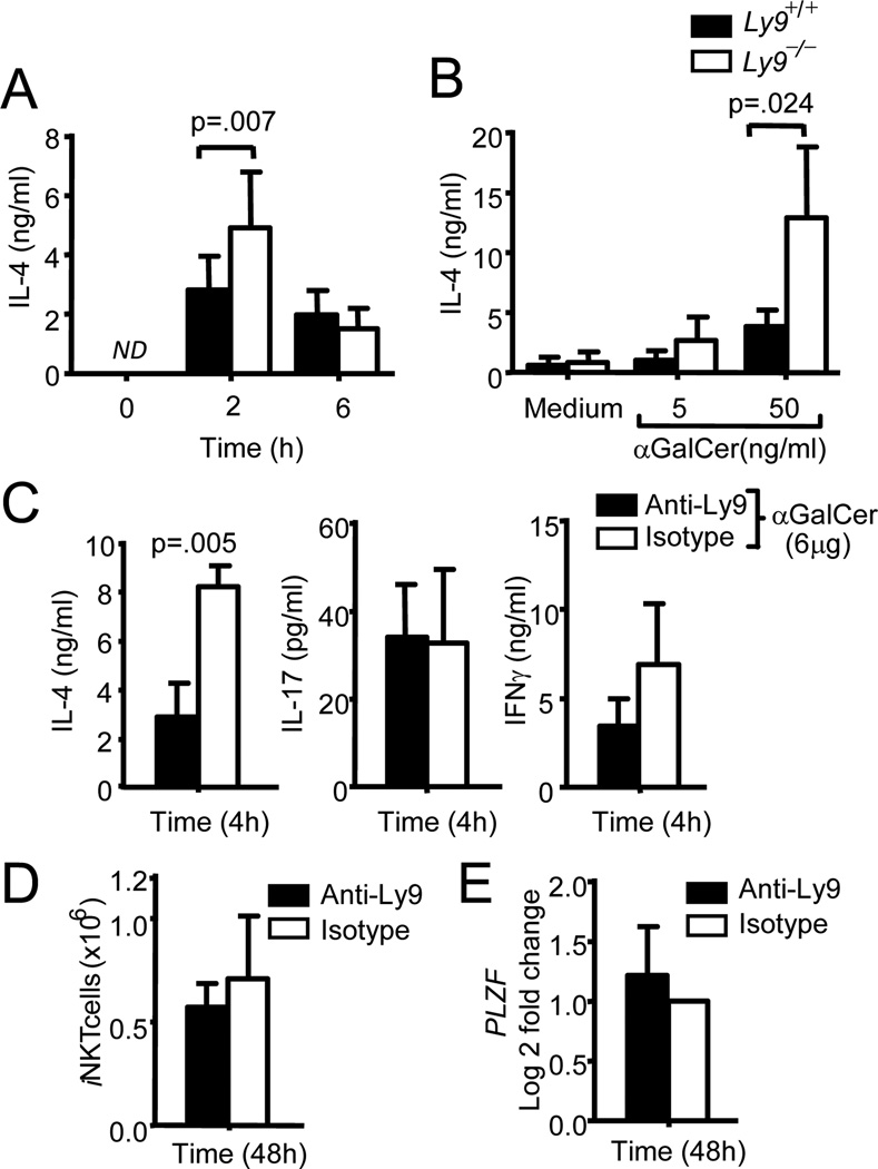
Figure 4. Ly9 deletion enhances cytokine production in iNKT stimulated cells.
(A) IL-4 in plasma from Ly9+/+ and Ly9−/− mice (n=11) i.p. injected with αGalCer (6µg). Blood samples were collected at the indicated time points. (B) IL-4 levels in the culture supernatants of thymocytes from Ly9+/+ and Ly9−/− (n=7) activated with αGalCer for 48h. (C) IL-4 (left panel), IL-17 (middle panel) and IFNγ (right panel) measured in plasma from Ly9+/+ mice injected with anti-Ly9 or a matching isotype (each group treatment n=12) 48 h before administration of αGalCer. Blood was obtained 4 h after αGalCer stimulation. (D)iNKT cell numbers or (E) changes of PLZF mRNA expression levels after anti-Ly9 or a matching isotype treatment for 48 hours previous to αGalCer administration. Data are representative of at least two independent experiments.

