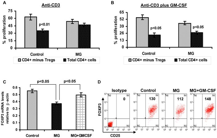
Figure 2.
In vitro GM-CSF treatment to lymphocytes enhances FOXP3 expression and suppressive function of Treg cells in MG. CFSE-labeled total CD4+ T cells and Treg cell–depleted CD4+ T cells were stimulated with human anti-CD3 alone or anti-CD3 plus GM-CSF in the presence of autologus irradiated APCs. Cultures consisting of total CD4+ cells (black bars) and CD4+ cells after removal of CD4+ CD25high CD127low/− cells (grey bars), were compared to evaluate suppressive function of Treg cells. After 5 days of culture, percentage of T cell proliferation was analyzed based on CFSE dilution. The bar diagram represents percentage Tresp cell proliferation in response to anti-CD3 alone (A) and anit-CD3 plus GM-CSF (B) for four MG patients and four healthy controls.(C) FOXP3 mRNA expression was determined by multiplex PCR using isolated CD4+ CD25high CD127low/−Treg cells obtained from a control subject, MG patient and an MG patient’s PBMCs treated with GM-CSF. Results are expressed as relative FOXP3 mRNA expression. Results are expressed as mean ± SEM. (D) Representative flow cytometry plots illustrate mean florescent intensity of FOXP3 expression within isolated CD4+ CD25high CD127low/− Treg cells.

