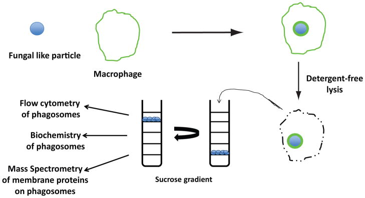
Figure 2.
Cartoon depicting method to isolate and analyze phagosomes containing fungal-like particles. Polystyrene beads with fungal-derived carbohydrates covalently attached to the surface are incubated with macrophages. After a defined period, detergent-free lysis releases cellular content including intact phagosomes. Lysates are added to discontinuous sucrose gradients and then are subjected to ultracentrifugation. Phagosomes are easily isolated as a result of the buoyant properties of polystyrene beads and then analyzed by flow cytometry, Western blot, or mass spectrometry of membrane proteins.

