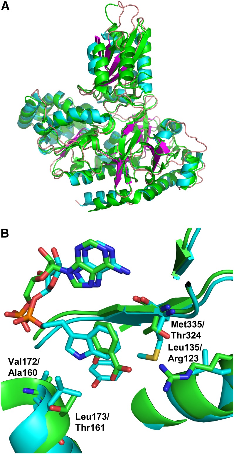
Figure 2.
Comparison of Vv GH3-1 to the At GH3-12 AMP-Bound Structure and the Salicylate-Bound Structure.
(A) Superposition of Vv GH3-1 with 4EPM (At GH3-12 with AMP). The root mean square deviation over 511 aligned residues (out of 554/565) is 1.3 Å with 49.5% sequence identity (as calculated with the SSM algorithm implemented in Coot). Vv GH3-1 is in cyan for α-helices, magenta for β-strands, and coral for the loops; At GH3-12 is in green.
(B) Superposition of Vv GH3-1 with 4EQL (At GH3-12 with SA and AMP). The root mean square deviation over 476 aligned residues (out of 494/565) is 1.3 Å (as calculated with the SSM algorithm implemented in Coot). GH3-1 is shown with cyan-colored carbon atoms (as above) with 4EQL in green. Specific residues are highlighted that are different between the two sequences and that may contribute to the different substrate specificities.

