Figure 3.
Time-Lapse Micrographs Illustrate Effects of Antibody Injection on Lily Pollen Tube Growth.
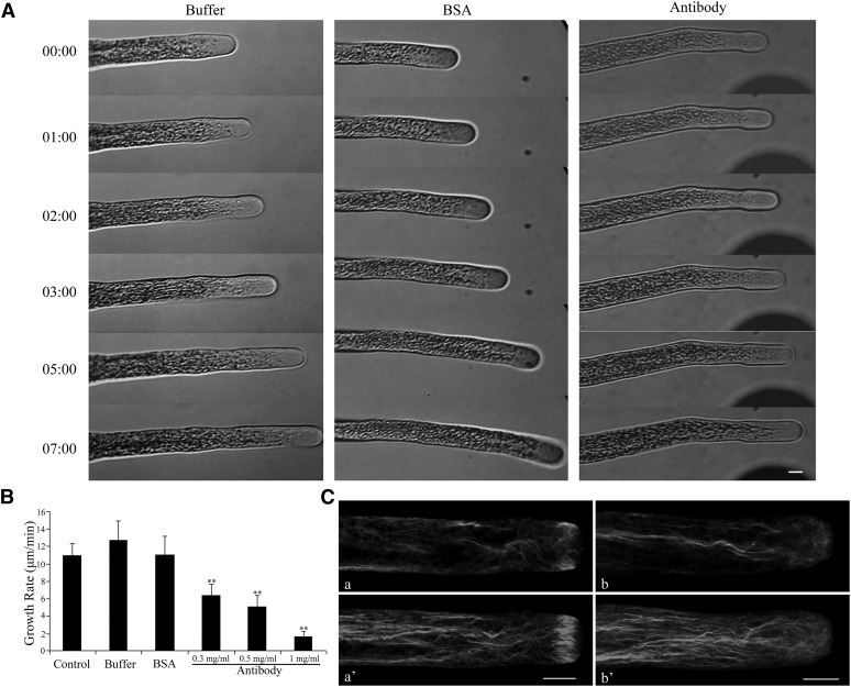
(A) Growing lily pollen tubes were microinjected with Ll-FIM1 antibody, BSA, or control buffer, as indicated. After 5 min of recovery, micropipette tips were slowly removed from the pollen tube, and the pollen tube was observed 10 min following injection. Time notated in minutes and seconds (mm:ss). Bar = 10 µm.
(B) The growth rate decreased significantly in the pollen tubes that were injected with antibody. Error bars represent mean ± se (**P < 0.01, by Student’s t test, n ≥ 15 per group).
(C) F-actin organization in antibody-injected pollen tubes and control pollen tubes. About 10 min after injection, cells were chemically fixed and stained with 200 nM Alexa-488 phalloidin. (a) and (a’) Control cell; (b) and (b’) cell injected with Ll-FIM1 antibody. All cells were visualized by confocal microscopy. (a) and (b) show medial confocal optical sections. The images in (a’) and (b’) are projections. Bar = 10 µm.

