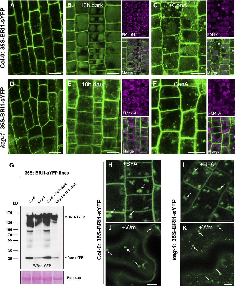
Figure 4.
KEG Regulates Vacuolar Transport of BRI1.
(A) and (D) Confocal microscopy images of root cells of wild-type (A) and keg-1 mutant (D) 35S:BRI1-sYFP transgenic seedlings grown under a 16-h/8-h light/dark cycle for 5 d and imaged near the end of the light cycle. Col-0, Columbia-0.
(B) and (E) The above seedlings were transferred to the dark ∼6 h into the light cycle on the sixth day and imaged 10 h later. These seedlings were also stained with the lipophilic dye FM4-64 to reveal vacuole structures.
(C) and (F) Seedlings treated with 0.5 μM ConA for 10 h.
(G) Total protein was extracted from 5-d-old 35S:BRI1-sYFP transgenic seedlings and immunoblotted with anti-GFP antibody. The red line indicates BRI1-sYFP degradation products.
(H) and (I) Seedlings treated with 100 μg/mL BFA for 1 h. Arrows indicate BFA compartments.
(J) and (K) Seedlings treated with 33 μM Wm for 1 h. Arrows indicate ring-like structures formed by MVB dilation.
Bars = 10 μm.

