Figure 2.
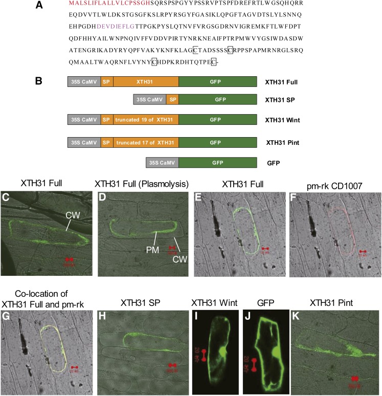
The Amino Acid Sequence of XTH31 and Its Subcellular Localization by Transient Expression in Onion Cells.
(A) The signal peptide at the N terminus, shown in red, is the putative wall/membrane targeting signal, the sequences in pink show the conserved GH16_XET domain, and conserved Cys residues (C) are in black squares.
(B) Schematic representation of XTH31 constructs fused to GFP. SP, signal peptide (19 amino acids at the N terminus of the full XTH31 coding sequence; see Methods for details). XTH31 Wint, the full XTH31 coding sequence (XTH31 Full) without the 19 putative SP residues; XTH31 Pint, the full XTH31 sequence without the first 17 of the 19 putative signal peptide residues. CaMV, Cauliflower mosaic virus.
(C) XTH31 Full-GFP localization. CW, cell wall.
(D) and (E) XTH31 Full-GFP localization in plasmolysed cells. PM, plasma membrane.
(F) pm-rk CD-1007 (plasma membrane marker) localization in the same plasmolysed cells as in (E).
(G) XTH31 Full-GFP colocalized with pm-rk CD-1007.
(H) XTH31 SP-GFP localization in plasmolysed cells.
(I) XTH31 Wint-GFP localization in nonplasmolysed cells.
(J) 35S:GFP localization in nonplasmolysed cells.
(K) XTH31 Pint-GFP localization in plasmolysed cells.
Red bars = 20 µm.

