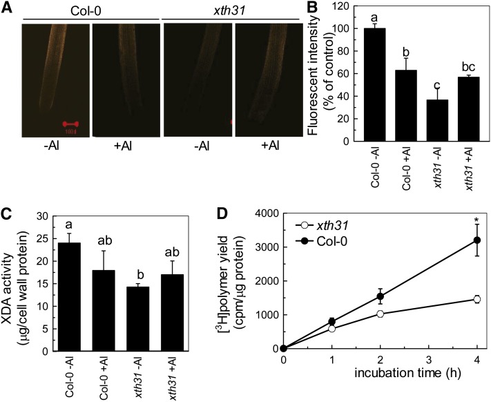
Figure 4.
Effect of Al on XET Action, Extractable XET Activity, and Extractable XDA in Col-0 and the xth31 Mutant.
Incubation with or without Al for 24 h corresponds to the panels “+Al” and “-Al,” respectively.
(A) Seedlings with ∼1-cm roots were grown on plates containing 0 or 50 µM Al for 24 h. Roots were then subjected to cytochemical assays of XET action for 1 h. Images reveal XET action as orange fluorescence in representative roots. Red bar = 100 µm.
(B) XET action expressed as fluorescence relative to untreated wild type. Data are means ± sd, n = 6. Different letters indicate significant differences at P < 0.05 by Student’s t test.
(C) Plants were treated with or without 50 µM Al for 24 h. Root extracts were subjected to in vitro assays of XDA. Data are means ± sd, n = 4. Different letters indicate significant differences at P < 0.05 by Student’s t test.
(D) Radiochemical assay of XET activity in root extracts. Buffer-extractable enzymes from the roots of Col-0 and xth31 seedlings were assayed for XET activity with tamarind xyloglucan and [3H]XXLGol as donor and acceptor substrates, respectively. Data are the mean ± se of six independent extracts. The asterisk shows a significant difference between xth31 and Col-0 at P < 0.05 by Student’s t test.

