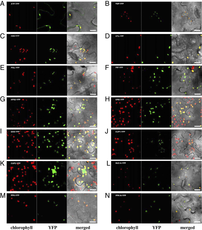
Figure 2.

YFP localization of selected candidate proteins. Tobacco leaves infiltrated with constructs in which the gene of interest was fused in front of YFP were analyzed by confocal laser scanning microscopy two days after infiltration. Chlorophyll autofluorescence is shown in the first channel and the YFP signal in the second channel. The third channel is a merged image of the previous two plus transmitted light. N after the name of a protein indicates that only its N-terminus was fused to YFP. Bar = 20 μm.
