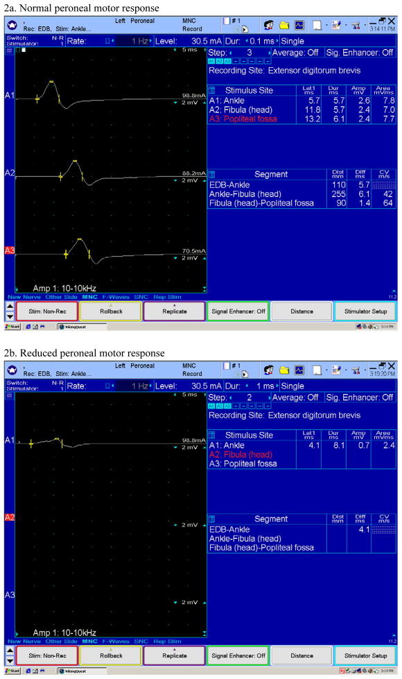
Figure 2.
Examples of normal and reduced peroneal motor responses obtained with surface electrodes by recording the extensor digitorum brevis muscle in the dorsum of the foot. In the reduced response (Fig. 2b) only a distal stimulation is performed because the response is very small. In the normal response (Fig. 2a) the peroneal nerve was stimulated at three different sites in the leg. Reduced motor responses may be seen in moderate to severe neuropathy.

