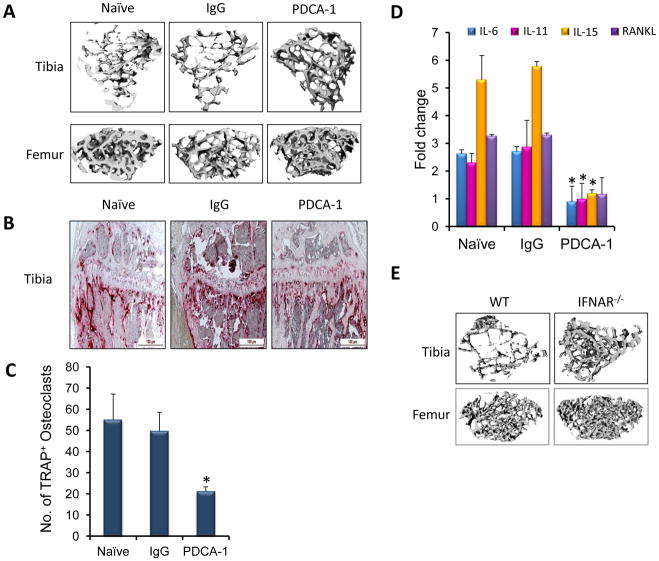
Figure 6. Decreased numbers of osteoclasts results in the absence of bone destruction in pDC depleted mice.
(a) Twelve days post-tumor challenge, tibia and femur from naïve, IgG-and PDCA-1-treated group were subjected to micro-CT analysis. (b) Paraffin sections of tibia from the naïve, IgG injected and PDCA-1 injected mice twelve days after tumor challenge were stained for TRAP to detect osteoclasts. (c) On day 12 post tumor challenge, monocytes and CD4+T cells from naïve, IgG and PDCA-1 antibody injected mice were co-cultured to assay osteoclast numbers. (d) cDNA from CD4+T cells of naïve, IgG- and PDCA-1 antibody treated mice were used to detect the levels of IL-6, IL-11, IL-15 and RANKL by real-time RT-PCR. Data is presented as a fold change in expression of RNA compared to the control. (e) Micro-CT analysis of tibia and femur from WT and IFNAR−/− mice 12 days post tumor challenge. For statistical analysis, N=3, *p<0.05 by Student’s t-test. Presented data is from 3 different mice from each group. These experiments were repeated twice separately.

