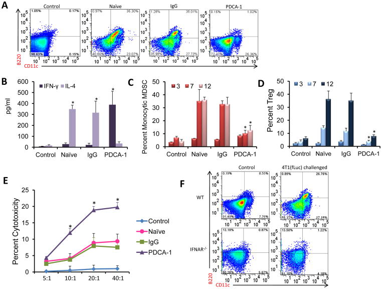
Figure 7. Depletion of pDC skews the immune response towards Th1 and results in decreased immunosuppression.
(a) Presence of pDC was detected in BM from naïve, IgG and PDCA-1 antibody injected mice 12 days post tumor challenge. A representative image from the 3 groups is presented. (b) IFN-γ and IL-4 levels were measured as mentioned before. (c) Numbers of monocytic MDSC (CD11b+Ly6C+Ly6Glo) in the BM after pDC depletion was enumerated by flow cytometry. (d) The presence of Treg cells was detected by staining BM cells from control, IgG and PDCA-1 antibody treated mice. (e) CD8+ T cells isolated from the BM of IgG and PDCA-1 treated animals 3 days post tumor challenge were used as the effector population (E) with 4T1(fLuc) cells as the target population (T). Cytotoxicity assay was carried out using a commercially available kit. (f) Presence of pDC was detected in BM from WT and IFNAR−/− mice 12 days post tumor challenge. Mice with no tumor challenge included as the control group. A representative image for each group, showing percent of pDC, is presented. Statistical analysis included N=3, *p<0.05 by Student’s t-test. The flow cytometry and cytotoxicity assays were from 3 mice for each group and for each time point. The experiments were repeated twice independently.

