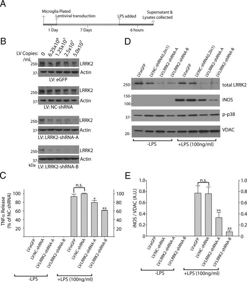
Figure 5.
LRRK2 knockdown attenuates inflammatory signaling in microglia. A, Graphical depiction of the experimental timeline used to generate lysates and serum from primary microglia cultures. B, A total of 5 × 10⁵ primary microglia per condition were exposed to the indicated copies of purified lentivirus encoding EGFP (no RNAi control), a noncoding control shRNA (NC-shRNA), or LRRK2 shRNAs (A, B), in 0.5 ml in culture. C, Serum from primary microglia treated with 2 × 10⁷ lentiviral copies/ml of the respective lentivirus for 7 d were analyzed for TNFα secretion 6 h post-LPS (100 ng/ml) or control exposures. Results are calculated from three independent experiments. D, Primary microglia exposed to LPS for 6 h as indicated were lysed in SDS buffer and analyzed by Western blot. Two micrograms of total protein was loaded per lane and analyzed with the indicated antibody. VDAC is used as a loading control. Blots shown are representative of three independent experiments. E, Quantification of iNOS levels normalized to VDAC expression. *p < 0.05, **p < 0.01 by one-way ANOVA with Tukey–Kramer, with respect to LV-NC-shRNA (+LPS) conditions. Error bars indicate SEM.

