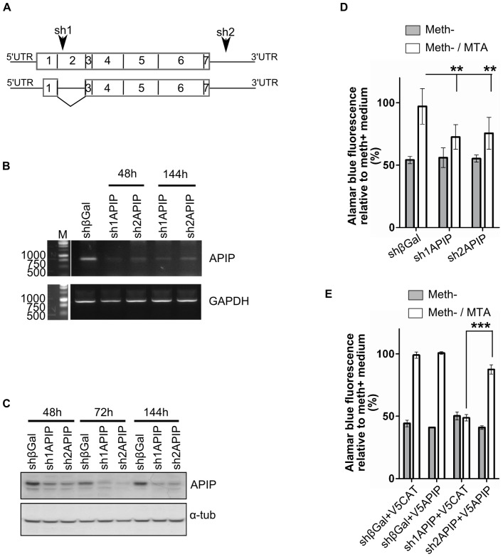
Figure 2. Transient silencing of APIP decreases the growth of the HeLa cells in MTA medium.
(A) Schematic representation of the two mRNAs isoforms of APIP. Sequence positions of the shRNAs used in the study (sh1APIP and sh2APIP) are indicated by arrows. (B) Semi-quantitative RT-PCR analysis of APIP silencing 48 h and 144 h after transfection with plasmids expressing shRNAs. A plasmid expressing shRNA against β-galactosidase (shβGal) was used as a negative control. The GAPDH gene was used as an internal control. (C) Western blot analysis of APIP silencing 48, 72 and 144 hours after transfection with plasmids expressing shRNAs. All lanes were loaded with 80 µg of cell lysate proteins. Anti α-tubulin (α-tub) was used as a loading control. The bands present below APIP were nonspecifically stained with the anti-APIP antibody. (D, E) Cell growth analysis of HeLa cells transiently silenced for APIP. Alamarblue fluorescence is expressed relative to the fluorescence of HeLa cells transfected with the same plasmids and cultured in normal methionine media. (D) 96 hours after transfection with plasmids expressing shRNAs, the same number of cells was grown for 48 hours either in complete media, or in methionine free media complemented or not with MTA, and their end point growth was measured by Alamarblue fluorescence (E) 72 hours after transfection with plasmids expressing shRNAs, cells were transfected with plasmids expressing the N-terminally V5-tagged APIP protein (V5APIP) or the N-terminally V5-tagged chloramphenicol acetyltransferase protein (V5CAT). 24 hours later, the same number of cells was grown for 48 hours either in complete media or in methionine free media complemented or not with MTA, and their end point growth was measured by Alamarblue fluorescence.

