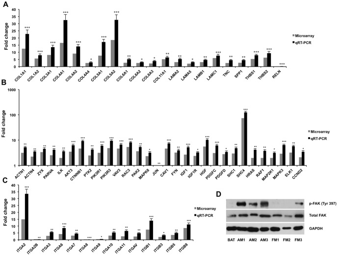
Figure 3. Expression of genes/proteins associated with the KEGG pathways in anaplastic meningiomas and arachnoidal tissue.
qRT-PCR experiments were performed to confirm the expression of genes in 6 anaplastic meningiomas (including the 3 tested samples in microarray) and 3 arachnoidal tissues. When expression of the genes in arachnoidal tissue was set to 1, relative expression of genes of ECM family (A) and integrins (C), as well as other genes (B) in the pathway is shown in each graph by using microarray and qRT-PCR. *P<0.05, **P<0.01, ***P<0.001. Western blotting determined total FAK and phosphorylated FAK (p-FAK Tyr 397) in anaplastic, fibroblastic meningiomas and arachnoidal tissue (D). GAPDH expression was used as controls. Key words: BAT, brain arachnoidal tissue; AM, anaplastic meningioma; FM, fibroblastic meningioma.

