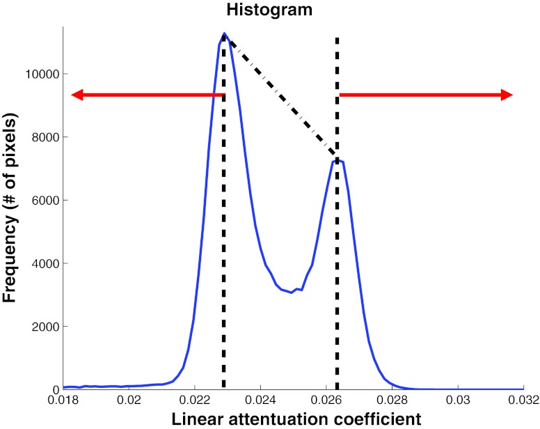
Figure 3.
Method used to convert the linear attenuation coefficient from the mastectomy specimen reconstruction to fibroglandular weight fraction. All voxels with linear attenuation coefficient less than the adipose peak were assigned 100% adipose tissue and those above the fibroglandular peak were assigned 100% fibroglandular tissue. Voxels with linear attenuation coefficient values between the two peaks were linearly scaled to be composed of a mixture of adipose and fibroglandular tissue.

