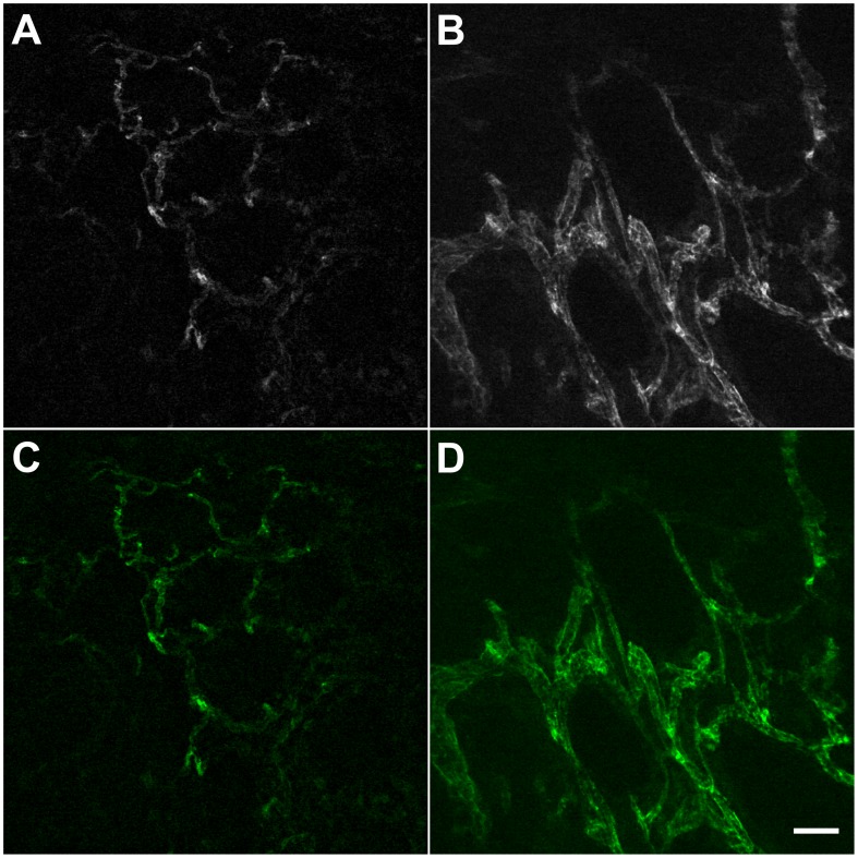
Figure 1. Ex-vivo CLE imaging with AF488-anti-CD31-antibodies.
In the normal mucosa, the vessels are organized in a hexagonal network (A); the corresponding area in the tumor shows more dilated, irregularly shaped vessels, with varying diameters along their length (B); the same optical sections are shown with added color overlay (C, D). *Scale bar is 50 µm.

