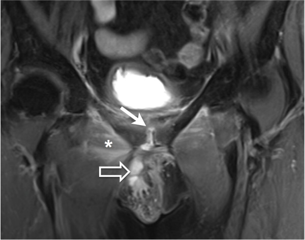
Figure 2.
Fat suppressed proton-density-weighted MRI sequence in the coronal plane shows hypersignal intensity on the pubic symphysis suggesting synovitis (arrow). These signal abnormalities seemed to be continuous with a voluminous abcess measuring 48 x 22 x 8 mm extending to the external genitalia (open arrow). Edematous infiltration of the right adductor (asterisk) is also shown.

