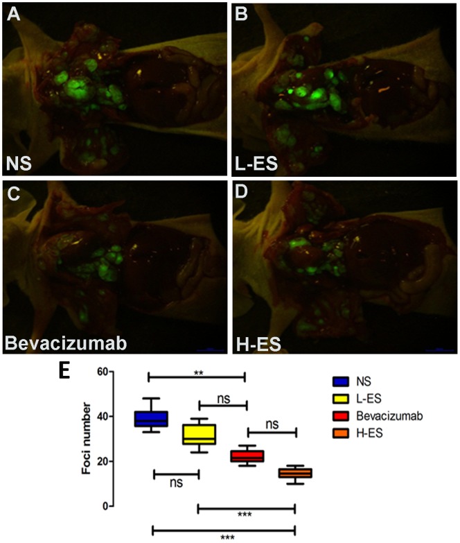
Figure 2. Tumor foci from four groups under fluorescence imaging system.
Mice were sacrificed and scanned by fluorescent imaging system. Fluorescent tumor foci were observed on the parietal and visceral pleura as well as hilar and mediastinal lymph node. The number of fluorescent pleural tumor loci was significantly decreased in H-ES group (D) compared with that in NS group (A) and L-ES group (B). The number of fluorescent pleural tumor loci in Bevacizumab group (C) was similar with that in H-ES group (D). (E): The difference of the number of Tumor foci on mice from four groups. Columns: mean value of each group, bars: ±SD. ***P<0.001, **P<0.01, *P<0.05. ns: no significant difference.

