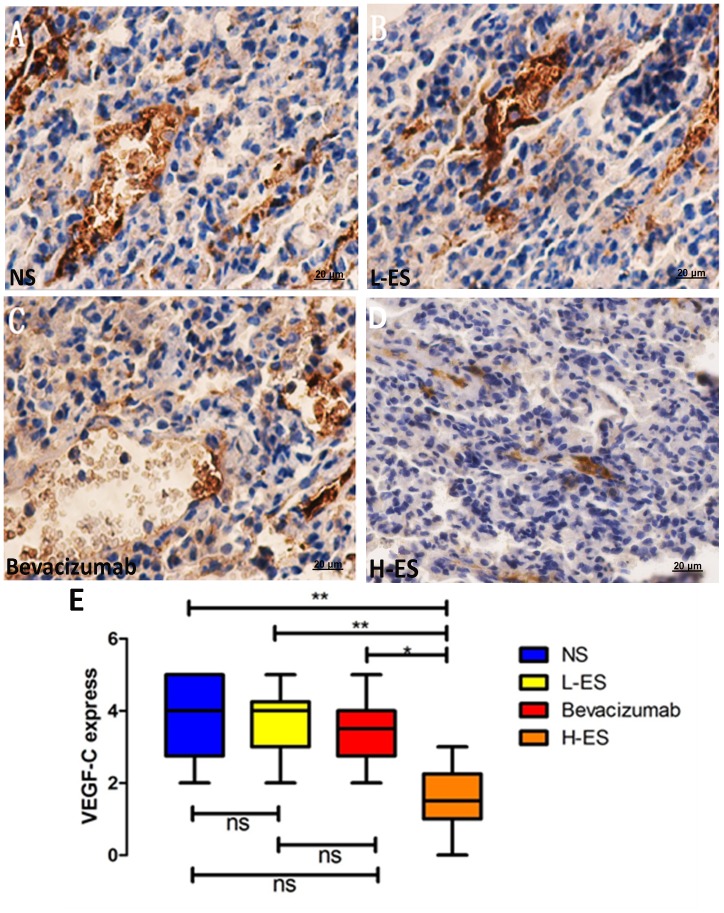
Figure 7. Immunohistochemistry staining of VEGF-C expression in the pleural tumors.
Positive immunohistochemistry staining of VEGF-C was shown as brown part in each figure. Expression of VEGF-C was accessed by the percentage of positive carcinoma cells and the staining intensity. The positive staining of VEGF-C in NS group (A) and L-ES group (B) indicated high expression of VEGF-C in these groups. Low expression of VEGF-C was shown in Bevacizumab group (C) and H-ES group (D). The expression of VEGF-C was significantly decreased in H-ES group compared with that in NS group or L-ES group or Bevacizumab group.Columns: mean value of each group, bars: ±SD. ***P<0.001, **P<0.01, *P<0.05. ns: no significant difference.

