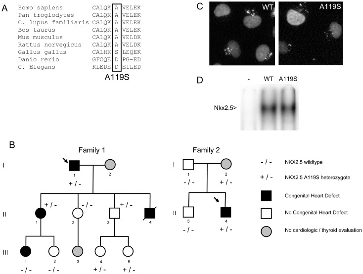
Figure 1. Family pedigrees, aminoacid alignments, nuclear localization of NKX2-5 protein and electro mobility shift assay.
A. Pedigrees of the two families with the p.A119S NKX2-5 mutation. Individuals with congenital heart defects are indicated with a filled black symbol, while individuals with normal echocardiography are indicated with a white symbol. Grey symbols represent individuals that have not been evaluated clinically. A slash denotes a deceased individual; the proband is indicated by an arrow. None of the evaluated family members showed thyroid abnormalities. Heterozygous carriers of p.A119S are represented by +/− and non-carriers by −/−. B. Multiple alignments of aminoacids of the region surrounding p.A119 for various species. C. Nuclear localization of either wildtype or p.A119S NKX2-5 protein in COS7 cells. Nuclei are stained in green, red represents either the wildtype or mutant protein, orange indicates nuclei that are positive for wildtype or mutant protein. D. Electro mobility shift assay in cos7 cells, using the published nkx2.5 binding site [26]. Wildtype nkx2.5 protein and p.A119S Nkx2.5 protein bind equally well, – is untransfected control.

