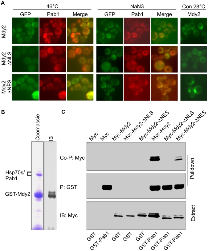
Figure 7. Mdy2 co-localize and interact with Pab1.
(A) Mdy2 co-localize with Pab1 following heat stress and treatment with sodium azide. GFP-Mdy2 and Pab1-RFP was visualized by fluorescence microscopy in a mdy2Δ strain transformed with plasmids containing GFP-Mdy2 (upper row), GFP-Mdy2-ΔNLS (middle row) or GFP-Mdy2-ΔNES (lower panel), and Pab1-RFP, after a temperature shift to 46°C (left panel) and after treatment with sodium azide (NaN3) (right panel). In the overlay pictures (merge), overlap of the colors appears yellow. GFP-Mdy2 and GFP-Mdy2-ΔNES but not GFP-Mdy2-ΔNLS are predominantly nuclear in control (Con) conditions at 28°C (right panel). (B) Mdy2 interacts with Pab1. Cell lysates from the GST-tagged Mdy2 strains were precipitated (P) with Glutathione Sepharose 4B. Following washing, the resin was eluted with glutathione. Eluted proteins were resolved by SDS-PAGE and visualized by immunobloting (control, IB) and Coomassie blue staining (Coomassie). Protein identities were established by mass spectrometry analysis. (C) Extracts from yeast strains HZH686 (W303-1A mdy2Δ) coexpressing GST alone (GST) or GST-tagged Pab1 (GST-Pab1) with Myc alone (Myc), Myc-tagged Mdy2 (Myc-Mdy2), Myc-tagged Mdy2-ΔNLS (Myc-Mdy2-ΔNLS) or Myc-tagged Mdy2-ΔNES (Myc-Mdy2-ΔNES) were subjected to pulldown using Glutathione Sepharose 4B as in Figure 4. The coprecipitation of indicated Myc-tagged Mdy2 proteins in the pulldown was confirmed by probing a Western blot with anti-Myc Ab (top panel, Co-P: Myc). To monitor pulldown recovery, the level of GST-Pab1 in the pulldown was measured by probing the same membrane with anti-GST Ab (second panel from the top, P: GST). Expression levels of indicated Myc-tagged Mdy2 proteins and GST-Pab1 in whole cell extracts (Extract) used for pulldown were measured on Western blots (third and fourth panels from top, IB:Myc and IB:GST, respectively).

