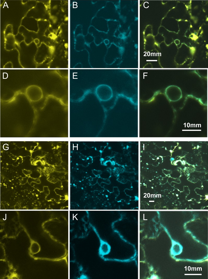
Figure 2.
Subcellular localization of AtADS1 and AtADS2 was investigated using transient expression in Nicotiana benthamiana leaves. A and D, Location of AtADS1-YFP; G and J, location of AtADS2-YFP; B, E, H, and K, distribution of ER marker CD3-953; C, merge of A and B; F, merge of D and E; I, merge of G and H; L, merge of J and K.

