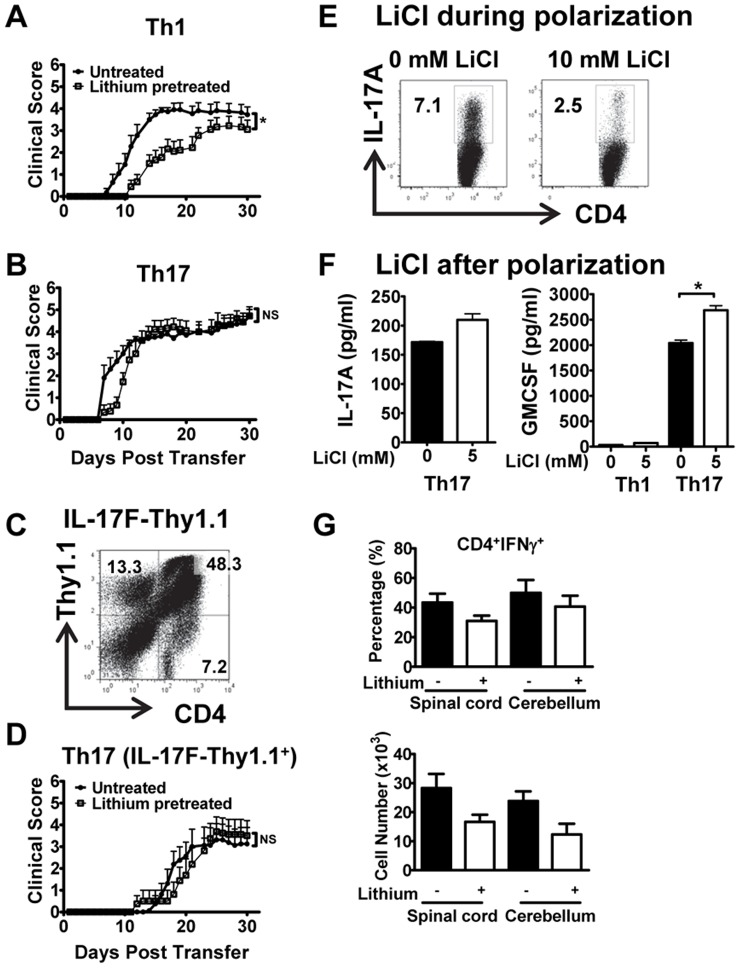
Figure 1. Lithium inhibits Th1-induced, but not Th17-induced EAE.
(A) Th1 cells (4–6×10⁶), (B) Th17 cells (4–6×10⁶), or (C and D) IL-17F-Thy1.1 cells (3–6×10⁵) were adoptively transferred i.v. into naïve untreated or lithium–treated recipient mice to induce EAE (mean ± SEM, n = 8–11 mice/group, *p<0.05 or NS, not significant, as determined by Mann-Whitney). (E) Intracellular cytokine staining for IL-17A in CD4+ gated cells from MOG35–55-immunized mice polarized to Th17 for 3 days in the absence or presence of 10 mM LiCl (representative dot plots and percentages are shown, n = 2). (F) 3 days polarized Th17 or Th1 cells from MOG35–55-immunized mice were cultured for an additional 24 h without or with addition of 5 mM LiCl, and production of IL-17A and GM-CSF was measured by ELISA in culture supernatants (one representative experiment is shown, n = 2; *p<0.05). (G) Infiltration of Th1 cells (CD4+IFN-γ+) into the spinal cord and cerebellum of untreated or lithium pretreated mice with adoptive transferred EAE. Infiltrating cells were isolated (14–15 d post transfer) and characterized by flow cytometry. Bar graphs depict percentage (top) and absolute number (bottom) of CD4+IFN-γ+ cells. Results are from 2–3 mice pooled per experiment (n = 2).

