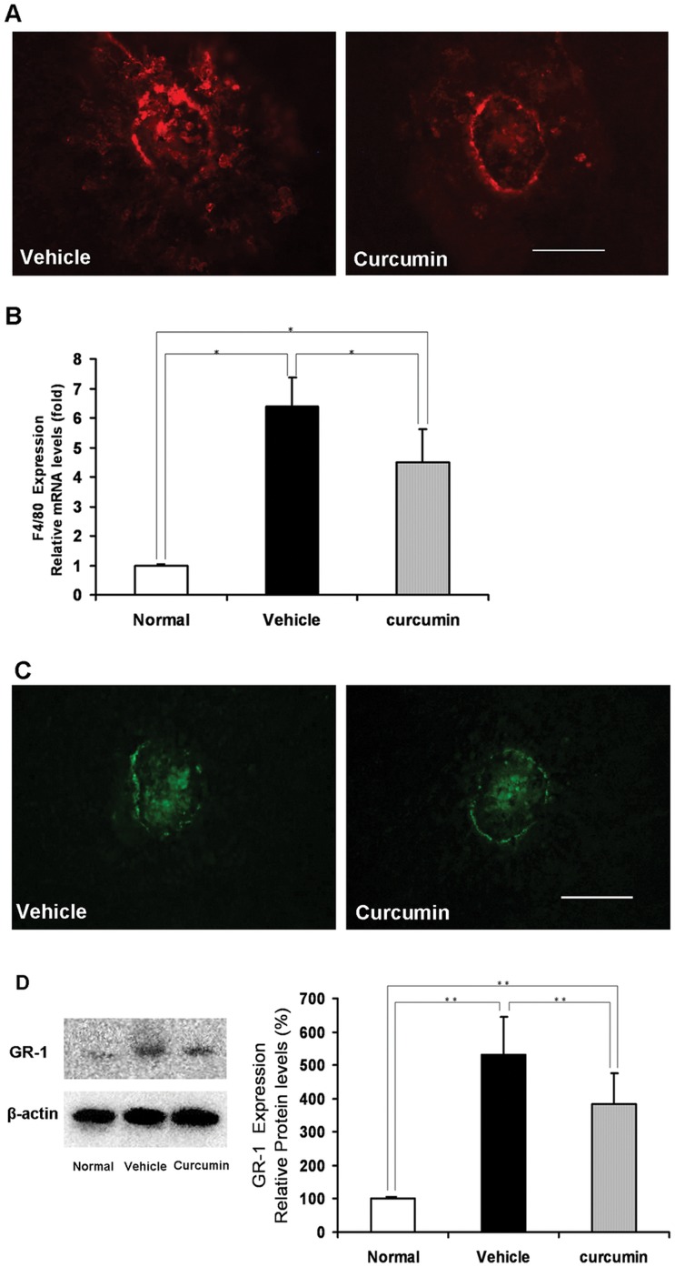
Figure 3. Inhibitory effect of curcumin on macrophages and granulocytes infiltration into CNV.
(A) Immunohistochemistry of macrophages (F4/80, red) in RPE–choroid flat mounts on day 3 (Scale bar = 100 µm). (B) The expression of F4/80 mRNA in RPE–choroid complexes on day 3 after photocoagulation. After photocoagulation, F4/80 mRNA expression significantly increased compared with no laser photocoagulation controls (relative to normal control). The increased F4/80 mRNA expression was significantly suppressed by curcumin treatment (n = 5, *P<0.05). (C) Immunohistochemistry of granulocytes (GR-1, green) in RPE–choroid flat mounts on day 3 (Scale bar = 100 µm). (D) Left: Representative Western blot showing GR-1 protein expression in samples from vehicle- and curcumin-treated mice on day 3 after photocoagulation. β-actin was used as a loading control. Right: Semi-quantitative analysis of the intensities of GR-1 bands from vehicle- and curcumin-treated mice. The mean for GR-1 in RPE–choroid complex of untreated mice was set at 100% (n = 5, **P<0.05).

