Fig. 3.
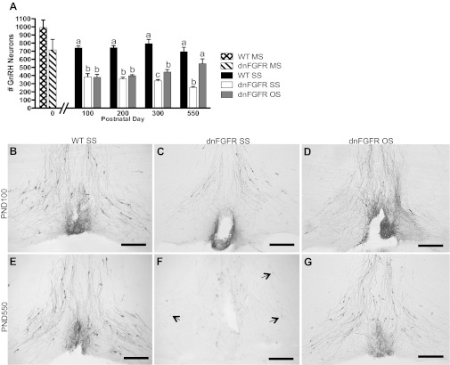
GnRH neuron number in OS- and SS-housed WT and dnFGFR males. A: GnRH neuron numbers were quantified in PND0 WT and dnFGFR male pups housed in mixed-sex (MS) configuration and in older WT and dnFGFR males that were SS or OS housed from PND30 to various ages. There were no differences between WT and dnFGFR MS pups at PND0 (n = 3–5/group; Student's t-test). From PND100 to PND550, dnFGFR SS males had significantly fewer GnRH neurons than WT SS males; OS in dnFGFR males significantly increased GnRH neuron numbers at PND300 and -550 but not earlier. Letter differences above bars represent significant differences (P < 0.05) within each age group (one-way ANOVA followed by Tukey's test; n = 4–7/group). B–G: representative photomicrographs of GnRH-immunoreactive (ir) neurons at the level of the organum vasculosum of the lamina terminalis (OVLT). At PND100, SS-housed WT males (B) had visibly more GnRH neurons than SS-housed (C) and OS-housed (D) dnFGFR males. At PND550, OS housing in dnFGFR males (G) visibly increased the number of GnRH neurons to a level higher than dnFGFR SS males (F) and comparable to WT SS males (E). Black arrows indicate lightly stained GnRH-ir soma in the SS-housed PND550 dnFGFR males (F). Scale bar = 200 μm.

