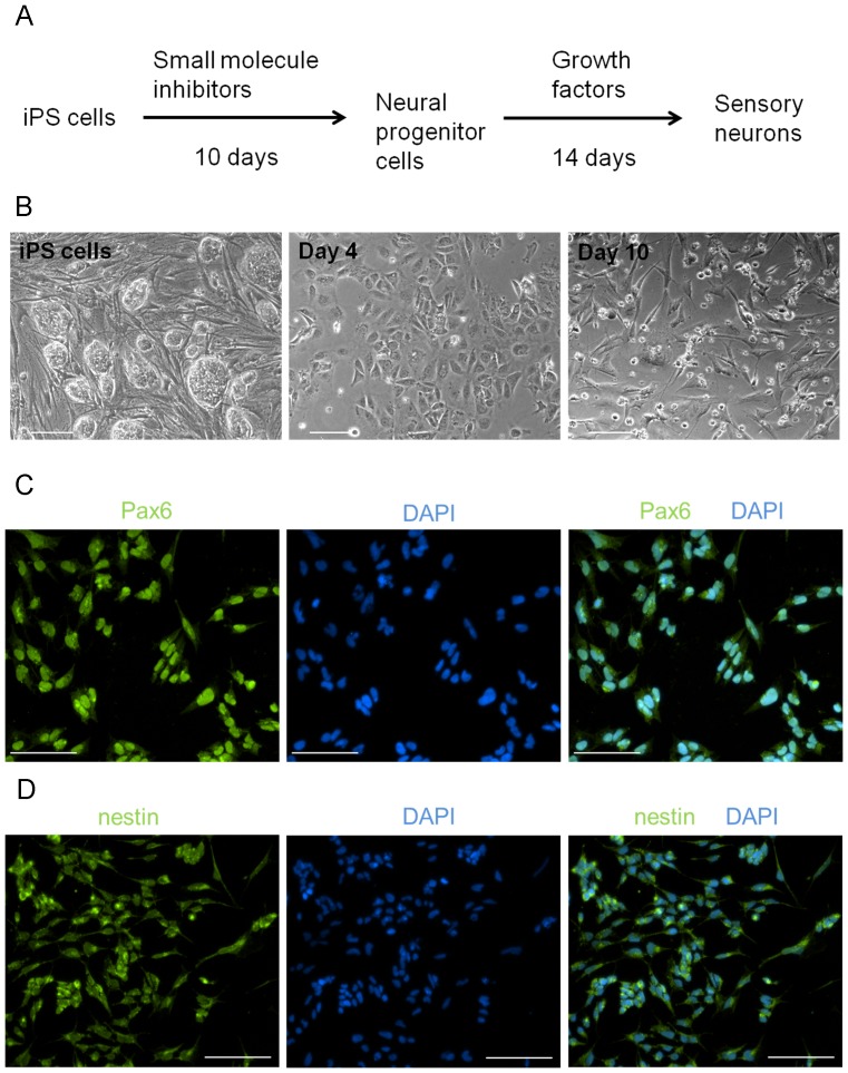
Figure 1. Conversion of human iPS cells to neural progenitor cells.
(A) Outline of the differentiation protocol. Human iPS cells were dissociated and plated on Matrigel-coated plates. For the first 10 days, they were exposed to small molecule inhibitors, followed by culturing for two weeks in growth factors. (B) Brightfield images of iPS cells after 4 and 10 days of exposure to small molecule inhibitors. After 10 days, iPS cells expressed (C) Pax6 and (D) nestin, markers of neural progenitor cells. Nuclei were visualized with DAPI. Scale for all images is 100 um.

