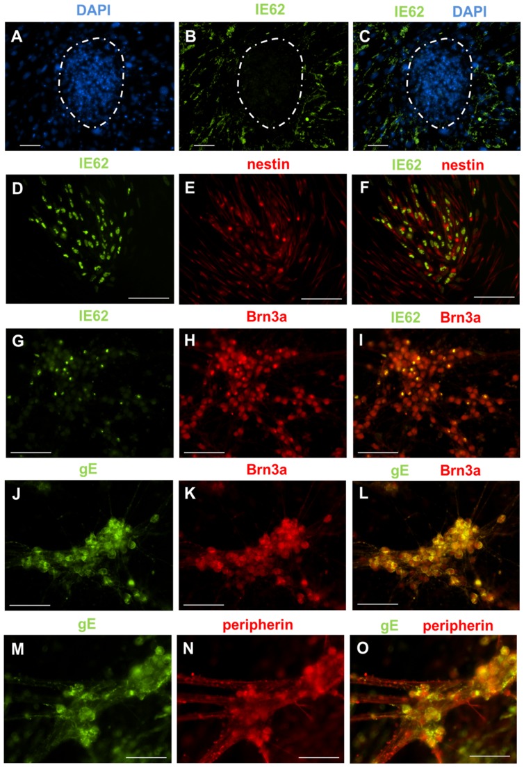
Figure 5. VZV infects iPS cell-derived neural progenitor cells and sensory neurons, but not undifferentiated iPS cells.
(A–C) Undifferentiated iPS cell cultures were infected with cell-free VZV at a MOI of 0.1 for 96 hours. iPS cells grow as colonies (dotted white circles) on a monolayer of non-dividing fibroblasts. (A) DAPI staining identifies the iPS cell colonies. (B) Immunostaining for IE62 revealed that only the monolayer of fibroblasts supported VZV infection. No IE62 staining could be observed in the iPS cell colonies. (C) Merge of panels A and B. (D–F) Neural progenitor cells infected with cell-free VZV at a MOI of 0.1 for 96 hours. Staining for (D) IE62 and (E) nestin. (F) Co-expression of IE62 and nestin indicated that neural progenitors cells can be infected by VZV (merge of panels D and E ). (G–O) Differentiated iPS cell cultures containing sensory neurons were infected with cell-free VZV at a MOI of 0.1 for 96 hours. Immunostaining for Brn3a (H, K), peripherin (N), IE62 (G) and gE (J, M) revealed that Brn3a+ cells co-expressed IE62 (I) and gE (L). Peripherin+ cells also co-expressed gE (O) and IE62 (data not shown). Collectively, these data show that sensory neurons can be infected by VZV. Scale for all images is 100 um.

