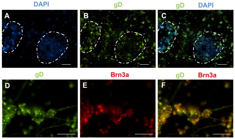
Figure 6. HSV infects iPS cells at all stages of differentiation.
Cells were infected with cell-free HSV at a MOI of 0.1 for 96 hours. (A) DAPI staining identifies the iPS cell colonies (highlighted by the dotted white lines). (B) Immunostaining for gD revealed that both iPS cells and the underlying fibroblasts supported HSV infection, as evidenced by abundant gD expression. (C) Merge of panels A and B. (D–F) Sensory neurons derived from iPS cells also supported HSV infection. Staining for gD (D) and Brn3a (E). (F) Merge of panels D and E. Scale for all images is 100 um.

