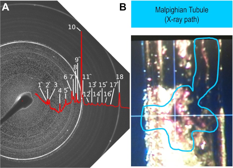
Fig. 5.
Crystal identity by X-ray diffraction of tubules. A: negative image and peak profile of X-ray diffraction pattern from Drosophilia Malpighian tubules after exposure to Na-oxalate (see materials and methods). Diffraction rings characteristic of crystal powders are annotated (1–18). B: single Malpighian tubule of 48-h high oxalate fed flies was placed in a quartz pipette for X-ray beam shooting (in blue line circle). Quartz does not disturb X-ray diffraction.

