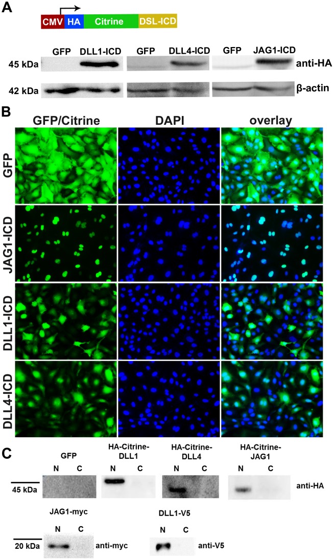
Figure 3. Localisation of Notch ligand intracellular domains.
(A) Scheme of the Notch ligand (DSL) intracellular domain (ICD) expression cassette in adenoviral vectors. Western blotting revealed expression of the fusion proteins in total lysates. (B) JAG1-ICD was localized predominantly in the nucleus, whereas DLL1-ICD and DLL4-ICD proteins were also localized in the cytoplasm. Scale bar, 100 µm. (C) Ligand-ICDs fused with Citrine were detected only in nuclear fractions (N) but not in the cytoplasmic fractions (C). HUVEC expressing full-length JAG1 or DLL1 fused to a c-terminal myc or V5-tag were lysed to obtain nuclear and cytoplasmic fractions. The processed intracellular fragments were detected only in the nuclear extracts by Western blotting.

