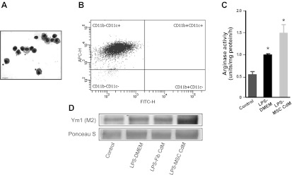
Fig. 4.
MSC-CdM promotes the M2 alveolar macrophage (AM) phenotype. A: representative photomicrograph of Hema3-stained AMs. Size bar: 15 μm. B: representative scatterplots of 4-color-stained AMs from experimental lungs. AMs were 98.2% CD11c+ and CD11b−. FITC-H, fluorescein isothiocyanate; APC-H, allophycocyanin. C: AMs exposed to LPS for 24 h had greater arginase activity compared with control AMs. MSC-CdM enhanced arginase activity compared with DMEM (n = 4/group). *P < 0.05 control vs. LPS-DMEM and LPS-MSC-CdM; LPS-DMEM vs. LPS-MSC-CdM. D: immunoblots of AM show enhanced induced Ym1 expression in LPS-MSC-CdM compared with LPS-DMEM and LPS-Fib-CdM (n = 4 samples/lane).

